Unlocking The Secrets: A Visual Journey Into Worms In Human Mouth
Worms in human mouth pictures refer to the visual documentation of parasitic worms that have infested the oral cavity of an individual. These images serve as crucial diagnostic tools for medical professionals, providing insights into the type and severity of the infestation, aiding in appropriate treatment planning.
The presence of worms in the mouth can stem from various factors, including poor oral hygiene, consumption of contaminated food or water, and underlying health conditions that compromise the immune system. Early detection and intervention are essential to prevent severe complications, such as tissue damage, nutrient deficiencies, and systemic infections.
Worms in human mouth pictures play a vital role in clinical practice by allowing healthcare providers to visualize the extent of the infestation, monitor treatment progress, and document the patient's condition for medical records and research purposes.
- Unveiling The Enigma The Mysterious Fate Of Clarence Thomass First Wife
- Unveiling Mysteries Elizabeth Youssefs Illness Explored
- Jenna Lyons Height Surprising Truths And Fashion Revelations
- Unveiling Whitney Wrens Net Worth A Journey To Discover Hidden Riches
- Unveiling The Visionary Impact Of Chris Carlos On Atlanta
Worms in Human Mouth Pictures
Worms in human mouth pictures, a visual representation of parasitic infestations in the oral cavity, play a crucial role in medical diagnosis and treatment. Here are eight key aspects related to this topic:
- Diagnostic Tool: Visualizing the type and extent of worm infestation.
- Treatment Planning: Guiding appropriate treatment strategies based on the severity of the infestation.
- Monitoring Progress: Tracking the effectiveness of treatment and assessing the patient's response.
- Medical Documentation: Providing visual evidence for medical records and research purposes.
- Public Health: Raising awareness about the causes and consequences of oral worm infestations.
- Educational Resource: Illustrating the lifecycle and behavior of parasitic worms in the oral cavity.
- Research Tool: Contributing to the study of oral parasitic infections and developing new diagnostic and treatment methods.
- Forensic Evidence: Assisting in the identification of parasitic infections in forensic investigations.
These aspects highlight the significance of worms in human mouth pictures in clinical practice, public health, and scientific research. They provide valuable insights into the diagnosis, treatment, and prevention of oral worm infestations, ultimately contributing to improved patient outcomes and overall health.
Diagnostic Tool
Worms in human mouth pictures serve as a vital diagnostic tool for medical professionals, enabling them to visualize the type and extent of worm infestation in the oral cavity. This information is crucial for accurate diagnosis and effective treatment planning.
- Uncovering Brendan Pennys Relationships Secrets And Revelations
- Unraveling The Mystery Ontario Brian Renaud Accident And Its Impact
- Unveiling The Secrets Of Sb19 Before Surgery Discoveries And Insights That Will Transform Your Surgical Journey
- Unraveling The Mystery Of Luis Miguels Mother Discoveries And Insights
- Unveiling The Life And Impact Of Andy Cohens Husband Discoveries And Insights
By examining worms in human mouth pictures, dentists and physicians can determine the species of worm involved, assess the severity of the infestation, and identify any associated tissue damage or inflammation. This visual evidence helps guide appropriate treatment strategies, such as the selection of specific antiparasitic medications or surgical interventions.
Real-life examples highlight the practical significance of this diagnostic tool. For instance, in cases of oral myiasis, a condition caused by the infestation of fly larvae in the mouth, worms in human mouth pictures can reveal the presence of maggots and their location within the oral cavity. This information enables prompt removal of the larvae and initiation of proper wound care, preventing further complications.
Overall, worms in human mouth pictures are an indispensable diagnostic tool for healthcare providers, aiding in the accurate assessment and management of oral worm infestations.
Treatment Planning
Worms in human mouth pictures play a crucial role in guiding appropriate treatment planning for oral worm infestations. By visualizing the type and extent of the infestation, healthcare providers can determine the most effective treatment approach based on the severity of the condition.
In cases of mild infestations, topical antiparasitic medications may be sufficient to eliminate the worms and prevent further spread. However, in severe infestations or when the worms have caused significant tissue damage, oral or systemic medications may be necessary. Worms in human mouth pictures help guide the selection of the appropriate medication and dosage, ensuring targeted treatment.
For instance, in cases of oral candidiasis, a fungal infection commonly known as thrush, worms in human mouth pictures can reveal the presence of Candida hyphae, indicating a more severe infection. This visual evidence supports the decision to prescribe antifungal medications with a broader spectrum of activity and a higher dosage to effectively combat the infection.
Overall, worms in human mouth pictures provide valuable information that empowers healthcare providers to tailor treatment strategies to the specific needs of each patient, optimizing treatment outcomes and minimizing the risk of complications.
Monitoring Progress
In the context of oral worm infestations, worms in human mouth pictures play a critical role in monitoring treatment progress and assessing the patient's response. By visualizing changes in the type, number, and behavior of worms over time, healthcare providers can evaluate the effectiveness of the prescribed treatment and make necessary adjustments to optimize outcomes.
For instance, in cases of parasitic infections caused by roundworms or hookworms, worms in human mouth pictures can reveal a reduction in worm burden and motility after treatment. This visual evidence indicates a positive response to the antiparasitic medication and supports the continuation of the current treatment plan.
Conversely, if worms in human mouth pictures show minimal or no change after treatment, it may suggest treatment failure or resistance. In such cases, healthcare providers can consider alternative treatment options or adjust the dosage and duration of the medication to improve efficacy.
Overall, worms in human mouth pictures provide a valuable tool for monitoring treatment progress and assessing the patient's response to oral worm infestations. By tracking changes in the visual presentation of worms, healthcare providers can make informed decisions about treatment strategies, ensuring optimal patient outcomes.
Medical Documentation
Worms in human mouth pictures serve as crucial medical documentation, providing visual evidence for accurate patient records and research purposes. These images document the type, extent, and progression of oral worm infestations, aiding in the diagnosis, treatment planning, and monitoring of patient outcomes.
By incorporating worms in human mouth pictures into medical records, healthcare providers can create a comprehensive visual representation of the patient's condition. This documentation facilitates effective communication between healthcare professionals involved in the patient's care, ensuring continuity of treatment and preventing misinterpretations or oversights.
Furthermore, worms in human mouth pictures are invaluable for research endeavors. They provide a visual database for studying the epidemiology, pathology, and treatment of oral worm infestations. Researchers can analyze these images to identify patterns, trends, and potential risk factors associated with these conditions, contributing to the advancement of medical knowledge and the development of improved diagnostic and therapeutic strategies.
In conclusion, the connection between "Medical Documentation: Providing visual evidence for medical records and research purposes." and "worms in human mouth pictures" is vital for comprehensive patient care and scientific research. These images provide essential documentation for accurate diagnosis, effective treatment planning, and ongoing monitoring of oral worm infestations, while also contributing to the advancement of medical knowledge through research.
Public Health
Worms in human mouth pictures play a vital role in raising public health awareness about the causes and consequences of oral worm infestations. These images provide irrefutable visual evidence of the impact of parasitic worms on oral health, highlighting the importance of preventive measures and early intervention.
- Education and Prevention: Worms in human mouth pictures can be used in public health campaigns and educational materials to illustrate the consequences of poor oral hygiene and the importance of regular dental checkups. These images can effectively convey the risks associated with oral worm infestations, promoting preventive behaviors and encouraging individuals to seek prompt medical attention if symptoms arise.
- Surveillance and Monitoring: Worms in human mouth pictures contribute to surveillance and monitoring efforts by providing data on the prevalence and distribution of oral worm infestations in different populations. This information is essential for public health officials to identify high-risk areas, allocate resources effectively, and develop targeted interventions to prevent and control oral worm infestations.
- Research and Advocacy: Worms in human mouth pictures serve as powerful tools in research and advocacy efforts aimed at improving oral health outcomes. These images can be used to support grant proposals, illustrate research findings, and advocate for increased funding and policy changes to address the burden of oral worm infestations.
- Collaboration and Partnerships: Worms in human mouth pictures facilitate collaboration and partnerships between public health organizations, healthcare providers, and community groups. These images can be shared and discussed to raise awareness, develop educational materials, and implement targeted interventions to combat oral worm infestations.
In conclusion, worms in human mouth pictures are a valuable resource for public health efforts to raise awareness about the causes and consequences of oral worm infestations. These images play a crucial role in educating the public, supporting surveillance and monitoring, facilitating research and advocacy, and fostering collaboration to improve oral health outcomes.
Educational Resource
Worms in human mouth pictures serve as invaluable educational resources, illustrating the lifecycle and behavior of parasitic worms in the oral cavity. These images provide a unique window into the complex world of oral parasites, enhancing our understanding of their biology and the diseases they cause.
- Visualizing the Lifecycle: Worms in human mouth pictures offer a visual representation of the lifecycle stages of parasitic worms, from eggs to larvae to adult worms. This visual information is crucial for understanding the transmission, development, and reproduction of these parasites, aiding in the development of effective control and prevention strategies.
- Observing Behavior Patterns: These images allow researchers and healthcare providers to observe the behavior patterns of parasitic worms in the oral cavity. By studying the movement, feeding habits, and interactions of worms, scientists can gain insights into their ecology, pathogenesis, and potential for transmission.
- Illustrating Disease Manifestations: Worms in human mouth pictures provide visual evidence of the clinical manifestations of oral worm infestations. These images can help healthcare providers identify and diagnose different types of oral parasitic infections, assess the severity of the infestation, and determine appropriate treatment options.
- Educating Patients and the Public: Worms in human mouth pictures can be used as educational tools to raise awareness about oral worm infestations. By sharing these images with patients and the public, healthcare professionals can inform them about the causes, symptoms, and preventive measures associated with these infections.
In conclusion, worms in human mouth pictures are indispensable educational resources for understanding the lifecycle, behavior, and disease manifestations of parasitic worms in the oral cavity. These images play a vital role in research, diagnosis, patient education, and public health efforts aimed at preventing and controlling oral worm infestations.
Research Tool
Worms in human mouth pictures serve as a crucial research tool, contributing to the study of oral parasitic infections and the development of new diagnostic and treatment methods. These images provide valuable insights into the biology, behavior, and clinical manifestations of oral parasites, aiding researchers and healthcare providers in advancing our understanding and management of these infections.
By studying worms in human mouth pictures, researchers can identify and characterize new species of oral parasites, investigate their lifecycle and transmission patterns, and assess their virulence and pathogenicity. This knowledge is essential for developing targeted diagnostic tools, such as molecular assays and immunological tests, to accurately identify and differentiate between different types of oral parasitic infections.
Furthermore, worms in human mouth pictures facilitate the evaluation of the efficacy of new and existing treatment strategies for oral parasitic infections. By observing changes in the morphology, behavior, and abundance of worms in response to different treatments, researchers can determine the effectiveness of various antiparasitic drugs, assess the development of drug resistance, and optimize treatment protocols.
In conclusion, the connection between "Research Tool: Contributing to the study of oral parasitic infections and developing new diagnostic and treatment methods." and "worms in human mouth pictures" is vital for advancing our understanding and management of oral parasitic infections. These images provide a unique window into the world of oral parasites, enabling researchers to study their biology, behavior, and response to treatment, ultimately leading to the development of more effective diagnostic tools and therapeutic interventions.
Forensic Evidence
Worms in human mouth pictures play a crucial role in forensic investigations as they can provide valuable evidence of parasitic infections in deceased individuals. In cases of suspected parasitic infections, forensic pathologists and odontologists examine worms in human mouth pictures to identify the type of parasite involved and determine its potential role in the individual's death.
By analyzing the morphology, size, and location of worms in human mouth pictures, forensic experts can differentiate between different species of parasites and assess the severity of the infection. This information is essential for determining the cause of death and identifying potential links to other individuals or geographic locations.
For example, in cases of suspected foodborne parasitic infections, worms in human mouth pictures can provide evidence of the type of food consumed and the source of the infection. This information can assist in tracing the origin of the contaminated food and preventing further outbreaks.
Additionally, worms in human mouth pictures can help identify cases of parasitic infections that may have been overlooked during the individual's life. By examining these images, forensic experts can uncover evidence of past or chronic parasitic infections that may have contributed to the individual's overall health status and potentially played a role in their death.
In conclusion, the connection between "Forensic Evidence: Assisting in the identification of parasitic infections in forensic investigations." and "worms in human mouth pictures" is significant as these images provide valuable clues for forensic experts to determine the cause of death and identify potential links to other individuals or geographic locations.
Frequently Asked Questions about Worms in Human Mouth Pictures
This FAQ section provides concise answers to common questions and misconceptions surrounding worms in human mouth pictures.
Question 1: What are worms in human mouth pictures?
Worms in human mouth pictures refer to visual representations of parasitic worms that have infested the oral cavity. These images are captured for diagnostic, treatment, and research purposes.
Question 2: How are worms in human mouth pictures used for diagnosis?
Worms in human mouth pictures aid in diagnosing the type and severity of parasitic infestations in the oral cavity. By examining these images, healthcare providers can determine the species of worm involved and assess the extent of the infestation.
Question 3: How do worms in human mouth pictures help guide treatment?
Worms in human mouth pictures assist in planning appropriate treatment strategies. The type and severity of the infestation, as visualized in these images, guide the selection of specific antiparasitic medications or surgical interventions.
Question 4: What is the role of worms in human mouth pictures in monitoring treatment progress?
Worms in human mouth pictures play a crucial role in monitoring the effectiveness of treatment. By tracking changes in the type, number, and behavior of worms over time, healthcare providers can evaluate the patient's response to treatment and make necessary adjustments.
Question 5: How are worms in human mouth pictures used in research?
Worms in human mouth pictures serve as valuable research tools. They provide visual data for studying the epidemiology, pathology, and treatment of oral worm infestations. Researchers can analyze these images to identify patterns, trends, and potential risk factors associated with these conditions.
Question 6: What are the ethical considerations related to worms in human mouth pictures?
Worms in human mouth pictures involve ethical considerations related to patient privacy and informed consent. Healthcare providers must obtain appropriate consent from patients before taking and using these images for diagnostic or research purposes.
In conclusion, worms in human mouth pictures are valuable tools for diagnosing, treating, monitoring, and researching oral worm infestations. They provide essential visual information that aids in patient management, scientific investigations, and public health initiatives.
Please note that the information provided in this FAQ section is for general knowledge and informational purposes only, and does not constitute medical advice. It is essential to consult with a qualified healthcare professional for any health concerns or before making any decisions related to your health or treatment.
Tips Related to "Worms in Human Mouth Pictures"
Worms in human mouth pictures provide valuable insights for diagnosis, treatment, and research of oral worm infestations. Here are some crucial tips to consider:
Tip 1: Accurate DiagnosisWorms in human mouth pictures facilitate precise diagnosis of the type and severity of oral worm infestations. By examining these images, healthcare professionals can identify the species of worm involved and assess the extent of the infestation, leading to appropriate treatment planning.Tip 2: Guided TreatmentWorms in human mouth pictures guide the selection of effective treatment strategies. The type and severity of the infestation, as visualized in these images, inform the choice of specific antiparasitic medications or surgical interventions, optimizing treatment outcomes.Tip 3: Monitoring Treatment ProgressWorms in human mouth pictures play a vital role in monitoring the effectiveness of treatment. Tracking changes in the type, number, and behavior of worms over time allows healthcare providers to evaluate the patient's response to treatment and make necessary adjustments, ensuring optimal outcomes.Tip 4: Research and DocumentationWorms in human mouth pictures serve as valuable research tools and medical documentation. They provide visual data for studying the epidemiology, pathology, and treatment of oral worm infestations. Additionally, these images contribute to medical records, facilitating effective communication and continuity of care.Tip 5: Public Health AwarenessWorms in human mouth pictures can raise public health awareness about the causes and consequences of oral worm infestations. These images can be used in educational campaigns and materials to illustrate the importance of preventive measures and early intervention, promoting oral health and well-being.Key Takeaways:Worms in human mouth pictures are essential diagnostic tools for oral worm infestations. These images guide appropriate treatment planning and monitoring. They contribute to research, medical documentation, and public health initiatives. Accurate interpretation and ethical considerations are crucial when using worms in human mouth pictures.In conclusion, worms in human mouth pictures are indispensable for comprehensive patient care, scientific investigations, and public health efforts related to oral worm infestations. By following these tips, healthcare professionals and researchers can harness the full potential of these images to improve oral health outcomes and advance our understanding of these conditions.Conclusion
Worms in human mouth pictures provide a unique and valuable perspective into the diagnosis, treatment, and study of oral worm infestations. These images capture the presence, severity, and characteristics of parasitic worms in the oral cavity, aiding healthcare professionals and researchers in various aspects of patient care and scientific investigations.
The exploration of "worms in human mouth pictures" within this article highlights the multifaceted role of these images in:- Facilitating accurate diagnosis of oral worm infestations- Guiding appropriate treatment planning and monitoring- Contributing to research on the epidemiology, pathology, and treatment of oral worm infestations- Enhancing medical documentation and communication- Raising public health awareness about the causes and consequences of oral worm infestations
As the understanding of oral worm infestations continues to evolve, "worms in human mouth pictures" will undoubtedly remain a crucial tool for advancing our knowledge and improving patient outcomes. By embracing the potential of these images and adhering to ethical considerations, we can harness their power to effectively combat oral worm infestations and promote oral health worldwide.- Unveiling The Reasons Behind Mariahs Departure From Married To Medicine A Journey Of Discovery
- Unveiling The Enigmatic Wicked Witch Of The East Discoveries And Insights
- Unveiling Natasha Yis Net Worth Unlocking Financial Insights
- Unraveling The Mystery Of Luis Miguels Mother Discoveries And Insights
- Unveiling The Strength And Resilience Of Paquita La Del Barrio

4000x3000px Worms 3028.4 KB 362831
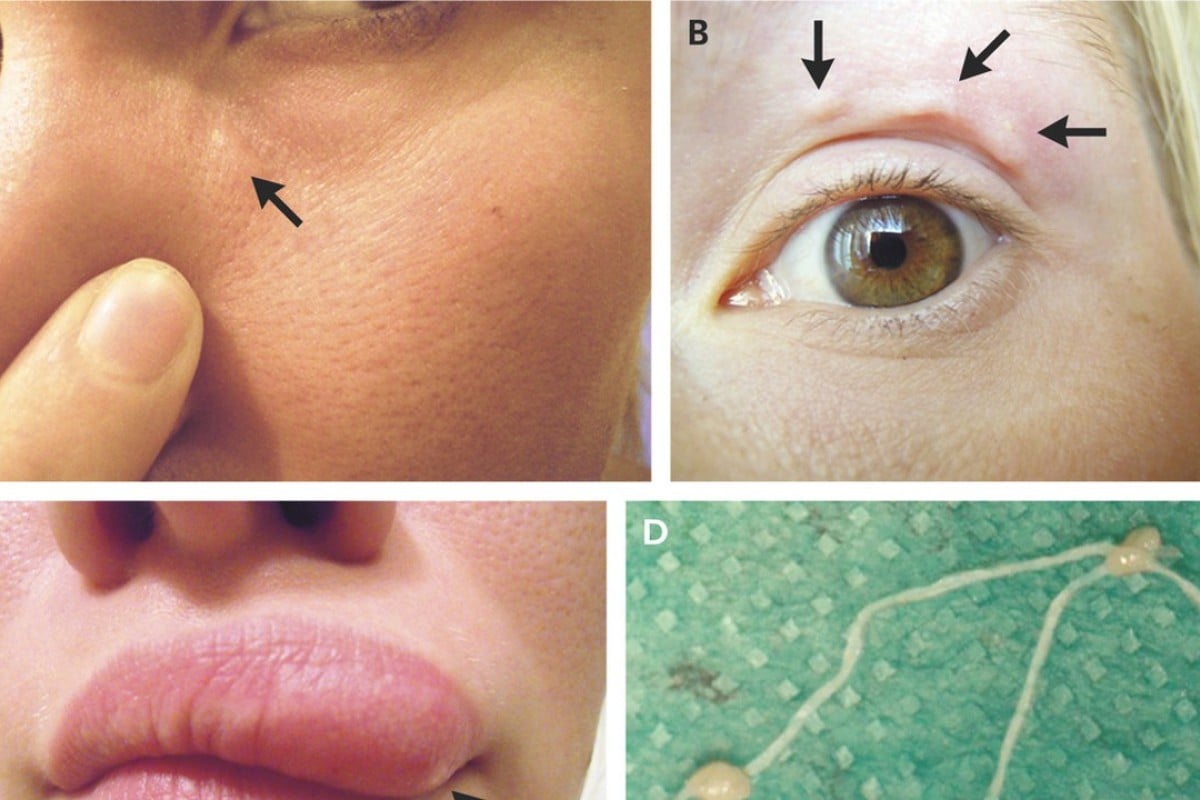
Those blemishes moving around on her face? Turns out it was a parasitic
